Indications
Pulmonary emboli, or blood clots in the lungs, are a very common and often debilitating problem. In 2008, the US Surgeon General and National Heart, Lung, and Blood institute issued a national call to action, declaring pulmonary emboli a major health threat. There are approximately 300,000-600,000 cases of new PEs/year resulting in 100,000-180,000 deaths. These blood clots most commonly originate from the veins in the legs and migrate up to the lungs. Fortunately, more often than not, they are small in size and can be treated with blood thinners. Approximately 25% of the time, however, these blood clots are large enough to cause a strain on the heart. Although patients may not feel different, the heart needs to work a lot harder to keep up with the stress of these blood clots. This is known as a sub-massive pulmonary embolus. Over the following hours to days, this stress may take such a toll on the heart, and medications may be required for the heart to function normally. This is known as a massive pulmonary embolus.
The traditional method of treating patients with pulmonary emboli has been with blood thinners. Research, however, has shown that patients who suffer from PE-related heart strain (submassive and massive PEs) are at risk for deteriorating. Equally as important, however, are the long term sequela of these PEs. This is known as chronic thromboembolic pulmonary hypertension, which occurs when the blood vessels in your lungs scar down from the blood clots. This results in a constant strain on your heart in order to pump enough blood to the lungs. In light of these potential problems, physicians have been treating patients with submassive and massive PEs with an aggressive clot-busting medication called Tissue Plasminogen Activator (TPA). This is known as systemic thrombolysis. Systemic thrombolysis involves injecting TPA through an IV anywhere in your body. TPA, however, carries a risk of causing bleeding elsewhere in the body. Physicians typically inject anywhere from 50-100mg of TPA during systemic thrombolysis.
In recent years, our physicians are able to use catheters placed in the pulmonary arteries to dissolve, break up, and remove blood clots from patients with submassive and massive PEs. This catheter-directed approach (known as pharmacomechanical catheter-directed thrombolysis or PMCDT) allows for much less TPA to be administered (typically 20 mg), which might reduce the risk of systemic bleeding. Even though the dose of medication is smaller, it is given directly into the clot, which can lead to improved results. By quickly treating the PE, this technique can rapidly reduce the amount of cardiac stress and can potentially prevent the clotted blood vessels from scarring over time. While giving a lot less TPA, typically 20 mg. This allows us to rapidly remove the stress your heart is under, and even more importantly, is able to prevent the clotted blood vessels from scarring over time.
Procedural Details
During the PMCDT procedure, our physicians insert a small catheter in a vein through a pinhole in the leg. Under live x-ray guidance, we are able to maneuver this catheter into the blood vessels of the lung that are harboring the clot. At this point, we typically infuse TPA at a slow and controlled rate over several hours. As opposed to giving 100 mg of TPA at once, we typically only infuse 2 mg of TPA per hour. During this infusion process, you are able to rest in bed, often overnight, within the ICU under the vigilant care of our pulmonary critical care specialists. After several hours, we bring you back to the angiography suite and inject x-ray dye through the catheter in order to document the resolution of the PE. If any emboli persist, we have several tools available to help break up the blood clots mechanically.
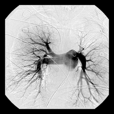
This image is from a pulmonary angiogram of the right lung, showing a PE at the beginning of the upper and lower lobe arteries.
This image is from a pulmonary angiogram of the right lung, showing improvement of the PE after PMCDT.
With the resolution of a majority of the clot burden, we can document the relieved strain on the heart. At this point, we remove the catheters and with the help of the pulmonary critical care team, help you in your transition home as soon as possible, typically within a few days.
Results
Studies have been performed to determine the outcomes following catheter-directed thrombolysis of PE in order to demonstrate that CDT can successfully treat patients with a submassive PE. The ULTIMA trial compared patients undergoing ultrasound-assisted catheter-directed thrombolysis with patients receiving only heparin for anticoagulation. This study showed that CDT improved right heart function faster than systemic anticoagulation. The SEATTLE II study showed that CDT successfully improved right heart function and pulmonary artery pressure. The PERFECT study showed a clinical success rate >80% in patients treated with CDT in addition to a significant reduction in pulmonary artery pressure. Other studies have performed as well. In 2016, Avgerinos, et al reported on their results comparing the use of catheter-directed thrombolysis with systemic anticoagulation. By comparing two groups of 64 patients undergoing each procedure, they demonstrated that catheter-directed thrombolysis is associated with a faster return of right ventricular function and a shorter stay in the ICU. However, more minor complications are seen with catheter-directed thrombolysis than systemic anticoagulation (usually minor bleeding). Both procedures otherwise reported similar outcomes.
Following the procedure, patients typically start a course of blood thinners for 3-6 months. This would have also been the case had you not chosen to go through PMCDT. The difference, however, is that you no longer have a significant amount of clots within the blood vessels supplying the lungs. Several studies have shown that your heart recovers to a baseline state much faster with PMCDT, most often within 24 hours. This results in markedly improved short-term morbidity and may theoretically translate to improved long-term morbidity. There is also a much lower risk of intracranial hemorrhage, or bleeding within the brain, in patients who have PMCDT compared to patients who receive systemic TPA without the use of catheters.

