Indications
Peripheral Arterial Disease (PAD) is a very common condition affecting up to 20% of Americans age 65 years and older. As of 2010, more than 200 million people worldwide and approximately 6% of Americans above the age of 40 are affected by PAD. It occurs due to atherosclerosis, or hardening of the arteries, within the pelvis and the legs. Atherosclerosis occurs when plaque, which is made of fat and cholesterol, builds up within the wall of the artery. This causes stenoses (narrowings) and occlusions (blockages) which limit the amount of blood that can flow through the arteries. There are several risk factors for PAD, including hypertension, high cholesterol, smoking, obesity, diabetes, inactivity, and a personal or family history of heart disease. It is always important for these conditions to be addressed with your primary care physician to reduce your risk of PAD.
Many patients with PAD have no symptoms. A common symptom is called intermittent claudication, which is muscle pain that typically occurs when walking and is relieved with rest. While this is a classic presentation, many patients present with atypical leg pain that often goes undiagnosed. These symptoms persist because the decreased blood flow to the legs can't keep up with the demand of muscles. When the blockages become very severe, the symptoms can include pain at rest, ulcers, and gangrene, which can possibly lead to loss of the affected limb. This is known as critical limb ischemia (CLI).
When a patient suspected with PAD presents to our office, we typically perform non-invasive tests to get an objective evaluation of the amount of blood flow to the legs. If your symptoms correspond with diminished blood flow, we proceed to make sure you are taking the necessary precautions to optimize perfusion to the legs. This includes medical optimization as well as life style changes. If this is not enough to address your symptoms, we then discuss the procedural treatment options for PAD.
Procedural Details
The hallmark of PAD treatment has always been revascularization. In other words, when patients are symptomatic with PAD, the solution to the problem is to find a way to increase blood flow to the legs. Historically, open surgery was the only option for revascularization. Due to recent strides in endovascular therapy pioneered by interventional radiology, the current expert consensus advocates for an “endovascular first” approach for nearly all patients suffering from PAD. As endovascular specialists, Albany IR is able to treat the stenoses and occlusions that result from PAD with minimally invasive cutting edge technology through a pinhole in the skin.
The day of your procedure, you arrive at our state-of-the art recovery room at Albany Medical Center. Shortly after, we bring you to our peripheral angiography suite. Once you are comfortable and sedated, we proceed by inserting a small catheter through a pinhole into an artery in your groin, the common femoral artery. We are able to advance this catheter into your aorta under live x-ray guidance. We then inject x-ray dye and get high-resolution diagnostic images of all of the major blood vessels proving flow to your legs. By obtaining a map of your vascular anatomy, we are able to pinpoint the exact blood vessels that are compromised with atherosclerotic plaque. We then advance a wire through the abnormal blood vessels we aim to treat. We are then able to slide the tools we use to fix the arteries over this wire, which essentially serves as a track for us to work over. Some of the commonly used tools include angioplasty balloons and stents. Angioplasty balloons are pre-sized balloons that we can use to dilate, or expand, stenotic blood vessels to a normal size. Stents are metal scaffolds that keep blood vessels open. With the diseased blood vessels fixed, we remove the catheter from the skin and you proceed to our recovery area where we monitor you for a few hours before going home. You are able to go home with nothing more than a band-aid.
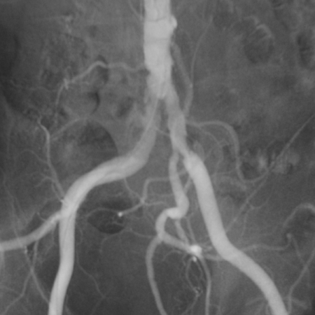
This image is from an angiogram of the abdomen and lower extremities. It demonstrates the aorta at the top of the image dividing into the common iliac arteries which bring blood to the legs. Stenoses (or areas of narrowing) are present at the origins of both of the common iliac arteries.

This image is from an angiogram of the abdomen and lower extremities after the placement of stents at the origins of the common iliac arteries. As a result of stent placement, more blood flow can now pass through these vessels to the legs, which can help ease symptoms.
Results
Generally speaking, patients with claudication or leg pain of any sorts experience immediate relief following the procedure. Patients with ulcers and wounds in their legs generally experience a much more rapid course of healing. Unless contraindicated because of other conditions, you are required to take a baby Aspirin daily in order to help keep the stents open. Following the procedure, we typically see you in our office at the 1 month mark. At that time, we obtain non-invasive vascular testing to ensure that perfusion to your legs is within normal limits. We then like to see you on a regular basis in order to ensure the stents remain patent, or open, and that you are not suffering from any symptoms of peripheral arterial disease.